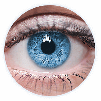
The doctors at VisionPoint Eye Center take special care to administer innovative treatments for your eye disease.
Amblyopia (Lazy Eye)
Lazy eye, medically known as amblyopia, is a loss or lack of development of vision, usually in one eye. This degenerative process usually begins with an inherited condition and appears during infancy or early childhood. Lazy eye needs to be diagnosed between birth and early school age since it is during this period that the brain “chooses” its visual pathway and may ignore the weaker eye permanently.
Lazy eye is not always easy to recognize since a child with worse vision in one eye does not necessarily have lazy eye. Because of this, it is recommended that all children, including those with no symptoms, have a comprehensive eye examination by the age of three and sooner if there is a family history of any eye condition or disease. If you suspect a problem, or need to set up your child’s first eye examination, contact VisionPoint Eye Center to set up an appointment.
Blepharitis
Blepharitis is a chronic infection of the eyelids. It often recurs and needs repeated treatment. Blepharitis is caused by inflammation of the eyelid margin, the edge of the eye from which your eyelashes grow. Debris and protein build-up at the base of eyelashes are called “collarettes” in blepharitis.
There are many factors that contribute to blepharitis. The most common factors are poor eyelid hygiene, blocked glands in the eyelids, excess oil produced by the glands in the eyelid, a bacterial infection (often staphylococcal) or an allergic reaction.
Careful, daily cleaning of the eyelashes can usually control blepharitis. This can be accomplished by moistening a clean washcloth with tap water as warm as you can stand without burning. Hold the washcloth against the eyelids until it cools, then re-warm and repeat for five to ten minutes. We recommend using a clock to follow the time. After soaking, each eyelid should be gently scrubbed for one minute.
If cleaning your eyelids at home isn’t alleviating your symptoms, your doctor may recommend BlephEx: a new, in-office procedure that allows your doctor to take an active role in cleaning your eyelid margins. This treatment cleans your eyelids and removes excess bacterial, biofilm, and bacterial toxins, the main causes of eyelid symptoms. With regular treatments, the eyelids stay clean and symptom-free. BlephEx is the first and only doctor eyelid cleaning procedure to help maintain clean and healthy lids.
The doctors at VisionPoint Eye Center can prescribe antibiotics if necessary. Blepharitis, if left untreated, can lead to severe infections. Always consult an eye care practitioner before beginning any treatment on your eyes.
Conjunctivitis
Conjunctivitis is an inflammation or infection of the conjunctiva, a thin, transparent layer covering the surface of the inner eyelid and the front of the eye. It affects people of all ages.
The three main types of conjunctivitis are infectious, allergic and chemical. The infectious form, commonly known as “pink eye,” is caused by a contagious virus or bacteria. Your body’s allergies to pollen, cosmetics, animals or fabrics often bring on allergic conjunctivitis. Irritants like air pollution, noxious fumes and chlorine in swimming pools may produce the chemical form.
If your eyes sting, itch or burn, you may be experiencing the common signs of “dry eye.” A feeling of something foreign within the eye or general discomfort may also signal dry eye.
If contagious, measures can be taken to prevent spreading conjunctivitis to others:
- Keep your hands away from your eyes
- Thoroughly wash hands before and after applying eye medications
- Do not share towels, washcloths, cosmetics, or eye drops with others
- Seek treatment promptly
Small children, who may forget these precautions, should be kept away from school, camp and the swimming pool until the condition is cured.
Certain forms of conjunctivitis can develop into a serious condition that may harm your vision. Therefore, it is important to have conjunctivitis diagnosed and treated quickly.
Torn and Detached Retina
A retinal detachment is a very serious problem that almost always causes blindness unless it is treated. The retina does not work when it is detached. Vision is blurred like a camera picture would be blurry if the film were loose inside the camera.
Causes of Retinal Detachment
The vitreous is a clear gel that fills the middle of the eye between the lens and the retina. As we get older, the vitreous may pull away from its attachment to the retina at the back of the eye. Usually the vitreous separates from the retina without causing any problems. But sometimes the vitreous pulls hard enough to tear the retina in one or more places. Fluid may pass through the retinal tear lifting the retina off the back of the eye, like wallpaper can peel off a wall.
The following conditions increase the chance that you might get a retinal detachment:
- Nearsightedness
- Previous cataract surgery
- Glaucoma
- Severe injury
- Previous retinal detachment in one eye
- Family history of retinal detachment
- Weak areas in your retina that can be seen by your eye doctor
Warning Signs of Retinal Detachment:
- Flashing lights
- New floaters
- A gray curtain moving across your field of vision
The doctors at VisionPoint Eye Center can diagnose retinal detachment during an eye exam where he or she dilates the pupils of your eyes. Some retinal detachments are found during routine eye exams. Only after careful examination can your eye doctor tell whether a retinal detachment is present.
Treatments for Tears and Detachments
Retinal Tears
Most retinal tears need to be treated with laser surgery or cryotherapy (freezing) to seal the retina to the back wall of the eye. These treatments cause little or no discomfort and may be performed in your eye doctor’s office. Treatment usually prevents retinal detachment.
Retinal Detachments
Almost all patients with retinal detachments require surgery to put the retina back in its proper place. Diabetic Retinopathy – If you have Diabetes Mellitus you are at risk of developing Diabetic Retinopathy. There are two types that can develop before you may notice any visual problems.
Droopy Eyelid
This condition can be onset at birth, develop with age or can happen as the result of an injury. Regardless of the situation that brought on droopy eyelids, a commonly used procedure called blepharoplasty can remove excess tissue from your upper and lower eyelids.
During blepharoplasty treatment, excess skin is removed from the eyelids to maintain the natural shape of the eyes and to restore a more youthful appearance to the face. In addition to the aesthetic benefits, blepharoplasty surgery can correct a condition in which sagging skin obscures vision, which may be covered by health insurance.
Flashes & Floaters
Flashes and Floaters are the warning signs for a more serious condition called a retinal detachment. Call and schedule an appointment with your eye doctor as soon as you notice any new flashes or floaters in your field of vision.
Floaters
Floaters are small specks or clouds that sometimes move in your field of vision. You can often see them when looking at a plain background like a blank wall or blue sky. Floaters are actually tiny clumps of gel or cells inside the vitreous, the clear jelly-like fluid that fills the inside of your eye.
While these objects look like they are in front of your eye they are actually floating inside. What you see are the shadows they cast on the retina, the nerve layer at the back of the eye that senses light and allows you to see. Floaters can have different shapes such as tiny dots, circles, lines, clouds or cobwebs.
The appearance of floaters may be alarming, particularly if they develop suddenly. You should see your eye doctor right away if you suddenly develop new floaters, especially if you are over 45 years of age.
Flashes
When the vitreous rubs or pulls on the retina, it creates a sensation of flashing lights. When the vitreous gel rubs or pulls on the retina, you may see what looks like flashing lights or lightning streaks. You may have experienced this same sensation if you have ever been hit in the eye and seen “stars”. As we grow older, it is more common to experience flashes.
Flashes of light can appear off and on for several weeks or months. However, as soon as you notice the sudden appearance of light flashes, visit your eye doctor immediately to see if the retina has been torn.
Causes of Floaters
When people reach middle age, the vitreous gel may start to thicken or shrink forming clumps or strands inside the eye. The vitreous gel then pulls away from the back wall of the eye causing a posterior vitreous detachment. It is a common cause of eye floaters.
Posterior vitreous detachment is more common for people who:
- even one new floater appears suddenly;
- you see sudden flashes of light; or
- you experience loss of side vision.
Treatment of Floaters
Floaters can get in the way of clear vision, which may be quite annoying, especially if you are trying to read. You can try moving your eyes, looking up and then down to move the floaters out of the way.
While some floaters may remain in your vision, many of them will fade over time and become less bothersome. When you notice new floaters you should have an eye exam immediately, even if you have had some floaters for years.
Floaters and flashes of light become more common as we grow older. While not all floaters and flashes are serious, you should always have a medical eye examination by your eye doctor to make sure there has been no damage to your retina.
Posterior Vitreous Detachment (PVD)
The center of the eye is filled with a jelly-like substance called the “vitreous”. At a young age, this jelly substance is very thick with a consistency somewhat like “jello”. With time, this jelly substance becomes more liquefied.
The vitreous is usually completely attached to the retina, which is the light sensitive seeing membrane in the back of the eye. As the vitreous jelly becomes progressively liquefied, it begins to move around inside the eye. Eventually, the vitreous becomes so loose that it “pulls away” from the retina behind it. This is called a “Posterior Vitreous Detachment (PVD).
Although a Vitreous detachment may occur at any age, most detachments occur over the age of 50. Studies show that almost 80% of the population experiences a Posterior Vitreous Detachment (PVD) by age 75.
Risk Factors for a Posterior Vitreous Detachment (PVD)
In general, a Posterior Vitreous separation is usually innocuous. Patients will typically experience the development of a large floater within their central vision. Although harmless, this is nevertheless annoying and may persist for several months.
Other risk factors include the more serious development of a retinal tear or detachment. This development would occur if the jelly substance teased and pulled away retina during separation. This may lead to a sight threatening condition known as a Retinal Detachment. Although uncommon, some literature suggests that a retinal tear may occur in 5%-10% of symptomatic patients during the first 6 weeks. For this reason, our doctors always perform a thorough dilated examination at the initial visit, and subsequent follow-ups. If the patient passes 3 months without incident they are typically followed annually.
Symptoms for PVD
- Onset of floaters or increase in number of floaters. (Floaters are often described as small black threads or cobwebs in vision.)
- Any association with flashes of white light in your peripheral or “side” vision, or an increase in flashes of light. These flashes would be more ominous if they occurred without exertion.
- A black curtain, or constant disturbance, noted in your side vision. You can check each eye by covering one eye at a time.
Refractive Errors
Astigmatism, Hyperopia, Myopia, and Presbyopia are all types of refractive errors that cause your eyes to see the way they do. They create the need for glasses or contacts to help your eyes process images clearly.
Astigmatism
Astigmatism is an uneven or irregular curvature of the cornea or lens, which results in blurred or distorted vision. Other symptoms of astigmatism include the need to squint, eye strain from squinting, headaches and eye fatigue.
In reality, most people have some degree of astigmatism, which is usually present at birth and is believed to be hereditary. In minor cases, treatment may not be required but is certainly beneficial. Moderate to severe astigmatism can be treated with corrective eyewear, contacts, or LASIK surgery.
Hyperopia (Farsightedness)
Farsightedness, medically known as hyperopia, refers to vision that is good at a distance but not at close range. Farsightedness occurs when the eyeball is shorter than normal, as measured from front to back, or when the cornea has too little curvature. This reduces the distance between the cornea and retina, causing light to converge behind the retina, rather than on it.
If you are mildly farsighted, your eye care provider may not recommend corrective treatment at all. However, if you are moderately or severely hyperopic, you may have several treatment options available, including eyeglasses, contacts, LASIK and photorefractive keratectomy (PRK). Your eye doctor will help you determine the best treatment option for you.
Myopia (Nearsightedness)
Nearsightedness, medically known as myopia, refers to vision that is good at close range but not at a distance. It generally occurs because the eyeball is too “long” as measured from front to back.
Nearsightedness is diagnosed during routine eye exams and possible treatments include eyeglasses, contacts, LASIK, and photorefractive keratotomy (PRK). Your eye care provider will suggest the best treatment option for you.
Presbyopia (Aging Eyes)
Aging eyes, medically known as presbyopia, is a condition in which the lens of the eye gradually loses its flexibility, making it harder to focus clearly on close objects such as printed words. Distance vision, on the other hand, is usually not affected.
Unfortunately, presbyopia is an inevitable part of aging and cannot be prevented by diet, lifestyle or visual habits. However, it is treatable with several types of corrective lenses, including progressives, bifocals and trifocals, single-vision reading glasses, multifocal contact lenses and monovision therapy.
Stye
A small area of redness and pain on the margin of your eyelid may indicate that you have a stye, known in medical terms as an external hordeolum. A stye is a blocked gland at the edge of the lid that has become infected by bacteria, usually Staphylococcus aureus.
The area of redness and pain will eventually form a ‘point’. Until this occurs, warm compresses should be applied to the area for 15 minutes three-to-four times a day. Our doctors often prescribe an antibiotic ointment in conjunction to warm compresses to aid in healing.
Remember that a stye that has not come to a ‘point’ should never be forcefully squeezed. Only when the stye has ‘pointed’, can it be gently expressed to empty its contents, after which the lids should be cleaned. Treatment with the ointment should be continued until symptoms have cleared. Sometimes it is necessary for the stye to be lanced to assist with expression.


